Monday, January 6, 2014

Other

-Acanthosis Nigricans-


-diffuse thickening and hyperpigmentation of the skin
-usually the axillae or body folds
-can be associated with hereditary, obesity, endocrine problems, meds, or malignancy
-caused by hyper secretion of pituitary peptide or non specific growth effect of hyperinsulinemia
-treatment is targeted at treating underlying disorder


-Burns-

-Rule of 9's-


-9% for the head and neck
-18% for the front torso
-18% for the back torso and buttocks
-9% for each lower extremity on front
-9% for each lower extremity on back portion
-9% for each arm
-1% for genitals


-Parkland Formula- to determine fluid needs for the first 24 hours in a burn victim

4 x weight in kg x TBSA burn = fluid requirements for the first 24 hours.

Give first half over the first 8 hours and give the second half over the last 16 hours.  Simple divide the total over the amount of hours necessary to give the fluids and that gives you the hourly rate.


-Classification of Burns-

-First degree Burn:


-Includes only the outer layer of skin, the epidermis
-Skin is usually red and very painful
-Equivalent to superficial sunburn without blisters
-Dry in appearance
-Healing occurs in 3-5 days, injured epithelium peels away from the healthy skin


-Second degree: Can be classified as partial or full thickness.


-Partial thickness
-Blisters can be present
-Involve the entire epidermis and upper layers of the dermis
-Wound will be pink, red in color, painful and wet appearing
-Wound will blanch when pressure is applied
-Should heal in several weeks (10-21 days) without grafting, scarring is usually minimal

-Full thickness
-Can be red or white in appearance, but will appear dry.
-Involves the destruction of the entire epidermis and most of the dermis
-Sensation can be present, but diminished
-Blanching is sluggish or absent
-Full thickness will most likely need excision & skin grafting to heal


-Third degree:


-All layers of the skin is destroyed
-Extend into the subcutaneous tissues
-Areas can appear, black or white and will be dry
-Can appear leathery in texture
-Will not blanch when pressure is applied
-No pain


-Fourth degree: Full thickness that extends into muscle and bone.


-Hidradenitis Suppurativa-


-chronic suppurativa often cicatricial disease of the apocrine gland axillae and the anogenital regain
-sometimes associated with nodulocystic acne and pilonidal sinuses
-unknown cause
-there is keratinous plugging of the apocrine duct, dilation of the hair follicle, and severe inflammatory changes of the single apocrine gland.
-Bacterial growth causes dilated duct
-ruptured duct or gland causes extension of inflammation or infection which cause tissue destruction and then this leads to ulceration, fibrosis and sinus tract formation
-treatment is incision and drainage and to excise recurrent fibrotic nodules and tracts


-Lipoma-


-benign subcutaneous tumors that are rounded, lobulated, and moveable over the overlying skin
-many are small but can be greater than 6 cm
-occur mainly on the neck, trunk and extremities
-most of the time just observe.  Rarely excise unless causing pain or discomfort


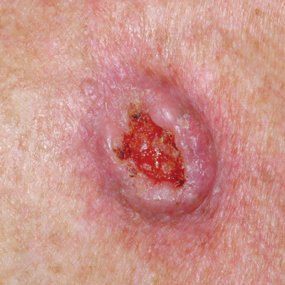
-Epithelial Inclusion Cyst-


-occurs secondary to traumatic implantation of the epidermis within the dermis.
-traumatically grafted epidermis grows in the dermis with accumulation of keratin within the cyst cavity
-treatment is excision


-Melasma-


-an acquired light or dark brown hyperpigmentation that occurs in the exposed areas to sunlight
-can be associated with pregnancy, oral contraceptives, or idiopathic
-pathogenesis is unknown
-treatment 3% hydroquinone solution in combination of tretinoin gel or 4% hydroquinone solution and glycolic acid
-need to use high SPF sunblock


-Pilonidal Disease-


-Pilonidal disease is a chronic infection of the skin in the region of the buttock crease
-The condition results from a reaction to hairs embedded in the skin, commonly occurring in the cleft between the buttocks.
-The disease is more common in men than women and frequently occurs between puberty and age 40. -It is also common in obese people and those with thick, stiff body hair.
-Treatment is incision and drainage with antibiotics.  Usually poly-microbial
-Definitive treatment is excision


-Pressure Ulcers-


-develop over body support surfaces over bony prominences as a result of the external compression of the skin, shear forces, or friction which produce ischemic changes or necrosis
-treatment is prevention.  Reposition patient every 2 hours.  Pad ulcer prone areas and massage them
-clean areas and keep free of urine and feces
-mobilize patient if possible

-Stages of Pressure Ulcers-
Stage I: Non-blanchable erythema
Intact skin with non-blanchable redness of a localized area usually over a bony prominence. Darkly pigmented skin may not have visible blanching; its color may differ from the surrounding area. The area may be painful, firm, soft, warmer or cooler as compared to adjacent tissue. Category I may be difficult to detect in individuals with dark skin tones.

Stage II: Partial thickness
Partial thickness loss of dermis presenting as a shallow open ulcer with a red pink wound bed, without slough. May also present as an intact or open/ruptured serum-filled or sero-sanginous filled blister.Presents as a shiny or dry shallow ulcer without slough or bruising. This category should not be used to describe skin tears, tape burns, incontinence associated dermatitis, maceration or excoriation.

Stage III: Full thickness skin loss
Full thickness tissue loss. Subcutaneous fat may be visible but bone, tendon or muscle are not exposed. Slough may be present but does not obscure the depth of tissue loss. May include undermining and tunneling. The depth of a Stage III pressure ulcer varies by anatomical location. The bridge of the nose, ear, occiput and malleolus do not have (adipose) subcutaneous tissue and Stage III ulcers can be shallow. In contrast, areas of significant adiposity can develop extremely deep Stage III pressure ulcers. Bone/tendon is not visible or directly palpable.

Stage IV: Full thickness tissue loss
Full thickness tissue loss with exposed bone, tendon or muscle. Slough or eschar may be present. Often includes undermining and tunneling. The depth of a Stage IV pressure ulcer varies by anatomical location. The bridge of the nose, ear, occiput and malleolus do not have (adipose) subcutaneous tissue and these ulcers can be shallow. Stage IV ulcers can extend into muscle and/or supporting structures (e.g., fascia, tendon or joint capsule) making osteomyelitis or osteitis likely to occur. Exposed bone/muscle is visible or directly palpable.


-Urticaria-


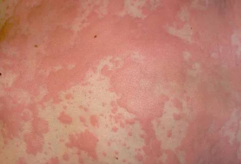
-IgE mediated complement mediated to physical stimuli
-acute urticaria is less than 30 days.
-chronic urticaria-greater than 30 days
-management is steroids, H1 and H2 blockers.  Subcutaneous epinephrine for hypotension, airway compromise.  Albuterol for bronchospasm.  Consider observation for severe systemic symptoms
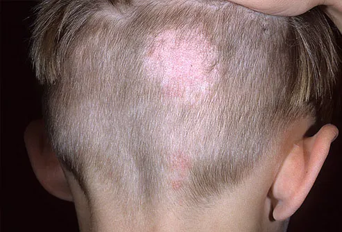

-Vitiligo-


-characterized by development of totally white macules and the complete absence of melanocytes
-associated with thyroid disease and many other medical conditions
-management sunscreens and cosmetic coverup
-Regimentation-  topical steroids, UVA, systemic UVA

Friday, January 3, 2014

Fungal Infections

Fungal Infections-

-Candidia-
-can be oral, genital, intertrigo, cutaneous , or balanoposthitis
-treatment dependent on site

-Oral Candidia-


-also called thrush
-get white creamy plaques on tongue or in mouth
-can be caused by dentures, oral inhaled steroids, diabetes, or immunosuppression
-common in infants
-treatment with nystatin swish and spit solution


-Vaginal Candidia-


-common in diabetics, recent antibiotic use
-diagnosed with KOH prep
-treatment with oral diflucan or anti fungal vaginal suppositories


-Balanoposthitis-


-transmitted in sexually active males by sexual contact
-causes burning itching and redness
-Azoles/imidazoles are more effective then nystatin
-can cause discrete pustules on the glans penis and the inner aspect of the foreskin
-can be mistaken for herpes


-Intertrigo Candidiasis-


-predisposing factors are obesity, diabetes, hyperhidrosis, heat
-initially pustules on erythematous base become eroded and confluent
-usually in the axillae, groin, perineal, inter-gluteal cleft
-treat by keeping area dry.  Topical treatment with topical anti fungal nystatin, and can use systemic oral anti fungal


-Cutaneous Candidia-


-vesicles, pustules, and papules become confluent in the folds
-can be treated with topical anti fungal or systemic for more severe cases


-Dermatophyte Infections-

-Tinea Pedis-


-dermatophyte infection of the feet
-causes redness, scaling, and maceration and or bullae formation
-often breaks up the integrity of the epidermis
-also known as athletes foot
-management use shoes while bathing.  Topical anti fungal for mild to moderate infections.  Oral anti fungal for more severe infection


-Tinea Manuum-


-dermatophyte infection of the hand.  Most commonly on the dominant hand
-Topical anti fungal for mild to moderate infections.  Oral anti fungal for more severe infection
-may see a raised well demarcated border


-Tinea Corporis-


-raised well demarcated border
-refers to a dermatophyte infection of the trunks, legs, arms, and neck
-Topical anti fungal for mild to moderate infections.  Oral anti fungal for more severe infection


-Tinea Capitis-


-Dermatophyte infection involving the hair follicles
-get a large circular area of missing patch of hair
-topical anti-fungals are ineffective
-oral anti-fungal necessary for several weeks.  Griseofulvin


-Tinea Barbae is a tinea infection of the beard

Bacterial Infections

Bacterial Infections

-Cellulitis-


-has similar features to erysipelas but extends to the deep subcutaneous tissues
-cellulitis lesions are not raised
-demarcation from uninvolved skin is distinct
-tissue is red, warm, and painful to palpation
-most common organism is staph aureus and group A streptococcus
-treatment parenteral-vancomycin and zosyn.  oral-cephalexin and bactrim or doxycycline


-Erysipelas-


-basically a superficial cellulitis with marked dermal lymphatic vessel involvement
-painful
-bright red, raised, edematous, indurated plaque with advance borders
-usually caused by group A beta hemolytic streptococcus
-can by caused by staph aureus
-treatment parenteral-vancomycin and zosyn.  oral-cephalexin and bactrim or doxycycline


-Impetigo-


-crusted golden yellow stuck on erosions that become confluent
-usually on nose, cheek, lips, and chin
-Caused by Group A Streptococcus
-Can be caused by staph
-Treatment usually with cephalexin, but should add bactoban, bactrim or doxycycline for MRSA coverage

Viral Diseases

Viral Diseases-

-Condyloma Acuminatum-


-may occur in the oral or genital epithelium
-HPV is the causative organism
-They can present from oral gentile contact and usually present flat papillomatous plaques or nodules with a glandular surface
-early lesions can be visualized by using 5% acetic acid
-lesions more advanced have a cauliflower appearance


-Exanthems-
-an infections generalized skin eruption associated with a primary systemic infection. 
-most common they are viral in nature, but can be associated with bacteria and parasites.  
-Examples of Exanthems-
-Rubella
-Measles
-Hand Foot and Mouth Disease
-Erythema Infectiosum


-Rubella-


-erythematous macules and papules appearing initially on the face and then inferiorly to the trunk and extremities
-Usually takes about 24 hours
-posterior auricular lymph node and posterior cervical lymph nodes can be enlarged
-see in patients not immunized


-Measles-


-highly contagious childhood viral infection
-presents with fever, coryza, cough, congestion, conjunctivitis, and Koplik spots
-see in patients that are not immunized
-caused by measles virus and paramyxovirus
-erythematous papules appear on the face and neck where they spread to the trunk and arms


-Hand Foot and Mouth Disease-


-multiple superficial erosions and small vesicles surrounded by erythematous halo.  
-lesions are painful
-occur on the hands, feet, and mouth (herpetic gingivostomatitis)
-caused mainly by cocksackie virus
-very contagious


-Erythema Infectiosum (Fifth's Disease)-


-caused by the Parvovirus
-get diffuse erythema and edema to the cheeks
-get a "slapped cheek" appearance
-prodrome of fever, malaise, headache, coryza, and sore throat
-management is symptomatic


-Herpes Simplex Virus-


-transmission is skin to skin, skin to mucosa
-presents as grouped vesicles on a erythematous base usually initially
-can have isolated lesions and varying degrees of ulceration
-HSV1-oral mucosal
-HSV2-genital
-Diagnosis is made my clinical suspicion confirmed with viral culture or antigen detection
-Treatment-acyclovir, valacyclovir, and famciclovir


-Molluscum Contagiosum-


-a self limited epidermal viral infection
-characterized by skin colored papules that are often umbilicated 
-can occur in children
-In HIV infected individuals can occur on the face 
-caused by poxvirus
-contagious can be transferred from skin to skin contact
-Aldara cream applied qHS 3 times a week for up to 1-3 months can help


-Varicella-


-highly contagious primary infection caused by the herpes zoster virus
-characterized by crops of pruritic vesicles that evolve to pustules, crusts, and ulcers.  
-the infection can have mild constitutional symptoms of fever, malaise and URI symptoms
-also called chicken pox
-acyclovir can shorten duration of illness of started early


-Herpes Zoster-
-is a dermatomal infection caused with reactivation of the varicella zoster virus characterized by unilateral pain in vesicular or bullous eruption limited to a dermatomal distribution
-major complication is post herpetic neuralgia
-acyclovir and antivirals can shorten duration of symptoms if started in the first 48 hours


-Verrucae-


-they are cutaneous human papilloma virus infections
-firm papules 1-10 mm are hyperkeratotic, cleft surface, and have vegetations 
-Verruca Plantaris (Plantar Warts)
-Verruca Plana (Flat Warts)
-can treat with salicylic acid or lactic acid
--Imiquimod cream can help
-Cryosurgery or electrosurgery for larger lesions


Thursday, January 2, 2014

Hair and Nails

Hair and Nails-

-Alopecia-


-Several different types-
-Alopecia Areata is considered an autoimmune disease.  Causes patches of hair loss over weeks to months.  Some may have spontaneous regrowth
-Alopecia Universalis is global loss of hair, eyebrows, lashes, beard and all body hair.
-Androgenic Alopecia-the most common progressive hairless that occurs through the combined effect of genetic predisposition and the action of androgen on the hair follicles of the scalp.
-Treatment for androgenic alopecia involves oral finasteride, topical minoxidil, and anti androgens


-Onychomycosis-


-chronic infection of the nail apparatus caused by dermatophytes most commonly.
-Also can be caused by candidia and molds
-80% occur on the feet
-a white patch is usually noted at distal nail.  Progressive infection the nail becomes opaque, thickened cracked, and yellow.  Raised by underlying hyperkeratotic debris in nail bed.
-Treatment involves debridement or systemic agents Terbinafine


-Paronychia-


-inflammation of the nail fold produces erythema, swelling, and throbbing pain and extend into the proximal nail fold and the eponychium
-purulent material accumulates and often requires surgical incision and drainage
-antimicrobial coverage with bactrim and cephalexin is necessary


Neoplasms

Neoplasms-

-Basal Cell Carcinoma-


-Four Clinical Types-
-Nodular
-Ulcerating
-Sclerosing
-Superficial Pigmented

-can be isolated but multiple lesions are not infrequent
-80% are on the head and neck
-excision is the goal.  Cryosurgery and electrosurgery are options.  Mohs surgery is sometimes needed
-Topical treatments can be used for superficial basal cell carcinomas but only for those that are below the neck.  
-Erivedge now available for metastatic basal cell carcinoma and locally recurrent basal cell carcinoma.  Once daily oral chemo agent
-Basal Cell Nevus Syndrome (BCNS) or Gorlin's Syndrome patients may have hundreds of basal cell lesions throughout their body


-Kaposi Sarcoma-


-multisystem vascular neoplasia 
-mucocutaneous violaceous lesions and edema and can involve any organ
-many individuals are immunocompromised, especially those with HIV
-lesions may be widespread or localized.  
-lesions almost always occur on feet, legs, or hands
-goal of management is control of symptoms of disease, not a cure.
-responds to radiotherapy of involved sites
-responds to systemic chemotherapy
-local therapy is usually directed at lesions that are individually cosmetically disruptive


-Melanoma-


-Lentigo Maligna Melanoma 
-Average onset is 45 greater in fair males
-Causes:  Sun burn and Heredity 
-Grows radial and extends deep only 0.5 mm 
-Makes up 5% of melanomas and has best prognosis 
-Lesions are dark in color with irregular border larger than an eraser head and flat 
-Seen in sun exposed areas 


-Superficial spreading melanoma
-70% of melanomas 
-Favors back, legs 
-Prognosis is fair because of a moderate radial growth phase 


-Nodular Melanoma 
-Grows deep 
-Worst kind 16% 
-Black bumps 
-Head neck and trunk favored 
-Metastatic Melanoma 
-A lesion with a blacked colored kidney shaped area in the lesion 

-Treatment-surgical excision is the goal.  May need concomitant chemo or radiation if metastatic


-Squamous Cell Carcinoma-


-Appears as a sharply demarcated scaling or hyperkeratotic macule, papule or plaque
-Solitary lesions are often pink with small erosions or crusted
-Increase incidence in males over 60 with fair complexion
-Keratinizing Epidermal cells
-Ultraviolet rays cause cancer.  HPV can also cause
-Sun exposed areas rolled border with crust in the center
-Can form cutaneous horn
-Surgical excision is the goal. Cryotherapy or 5 FU topically can also be used